Category Archives: Latest Research
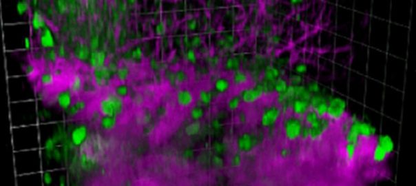
Multi-color 3-photon deep imaging is published in Science Advances
https://www.science.org/doi/10.1126/sciadv.abf3531
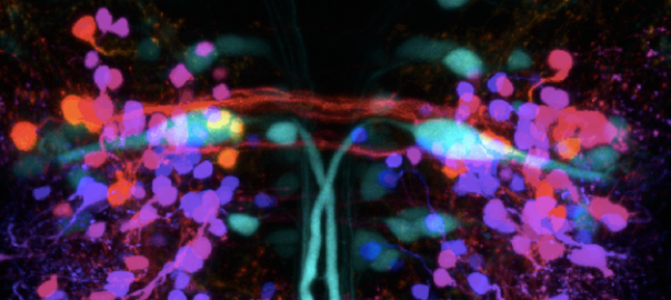
New imaging technique sheds light on adult zebrafish brain
Cornell scientists have developed a new technique for imaging a zebrafish’s brain at all stages of its development, which could have implications for the study of human brain disorders, including autism. See full Cornell Chronicle article →
Gerald Rubin to speak at Cornell Neurotech seminar, Sept. 24, 2021 4PM
The annual Cornell Neurotech Mong Family Foundation Seminar will feature Gerald M. Rubin. The seminar will be held in G10 Biotechnology on Sept. 23, 2021 at 4pm. The seminar title will be: Generating and using wiring diagrams of brain: Lessons from the Drosophila connectome Gerald Rubin is a Senior Group Leader at the Howard Hughes … Continue reading Gerald Rubin to speak at Cornell Neurotech seminar, Sept. 24, 2021 4PM
NeuroNex—a Radical Collaboration
An engineer and a neuroscientist gathered a group of Cornell scientists and engineers to tackle a frontier of science—the brain. Now, they form a hub. by Jackie Swift, CornellResearch Read the article and learn more here.
$9M grant will create neurotech research hub at Cornell

3-photon imaging of mouse brain activity is published in Nature Methods
Dimitre G Ouzounov, Tianyu Wang, et al. record spontaneous activity from up to 150 neurons in the hippocampal stratum pyramidale at ~1-mm depth within an intact mouse brain. Using three-photon microscopy at 1,300-nm excitation, the authors of a recently published Nature Methods paper, demonstrated that functional imaging of GCaMP6s-labeled neurons can be achieved beyond … Continue reading 3-photon imaging of mouse brain activity is published in Nature Methods

Revealing a circuit in the brain responsible for a behavioral choice
Brains are used to make behavioral choices about what to do next. Animals with two sides, including humans, constantly make choices about whether to respond to the left or right. Do they look left, or look right; turn left or right; reach left or right? Surprisingly, we know little about the wiring of the nerve … Continue reading Revealing a circuit in the brain responsible for a behavioral choice

Novel imaging of the degeneration underlying illnesses such as multiple sclerosis to allow for faster development of treatment strategies
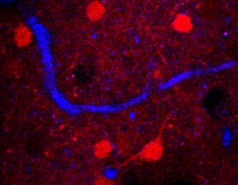
Normal brain function depends on wrappings of nerve cells by myelin which enhances the speed of conduction of electrical information in the brain and spinal cord. Disruptions of myelin are the source of the devastating movement problems with vision and movement in multiple sclerosis. This breakthrough allows the visualization of myelin, even on single nerve … Continue reading Novel imaging of the degeneration underlying illnesses such as multiple sclerosis to allow for faster development of treatment strategies

A new tool probes the inner workings of the brain
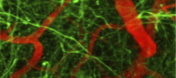
The application of three-photon microscopy allows for the visualization of the normal structure and function of single neurons deep in the living brain of mice, one of the most important model animals in neuroscience. Watching structure and function over time is needed to reveal how the brain works and what changes during disease. http://www.news.cornell.edu/stories/2013/01/three-photon-microscopy-improves-biological-imaging myelin … Continue reading A new tool probes the inner workings of the brain
Watching re-growing neurons inside the spinal cord to directly test treatments for spinal injury
Chronic imaging paperLooking deeply into living spinal cord of an animal over months will allow one to literally watch the efficacy of treatments for spinal cord injury. It also opens the possibility of watching the activity of neuronal circuits that generated movement in normal animals and after treatment. http://www.nature.com/nmeth/journal/v9/n3/abs/nmeth.1856.html Chronic imaging paper pdf