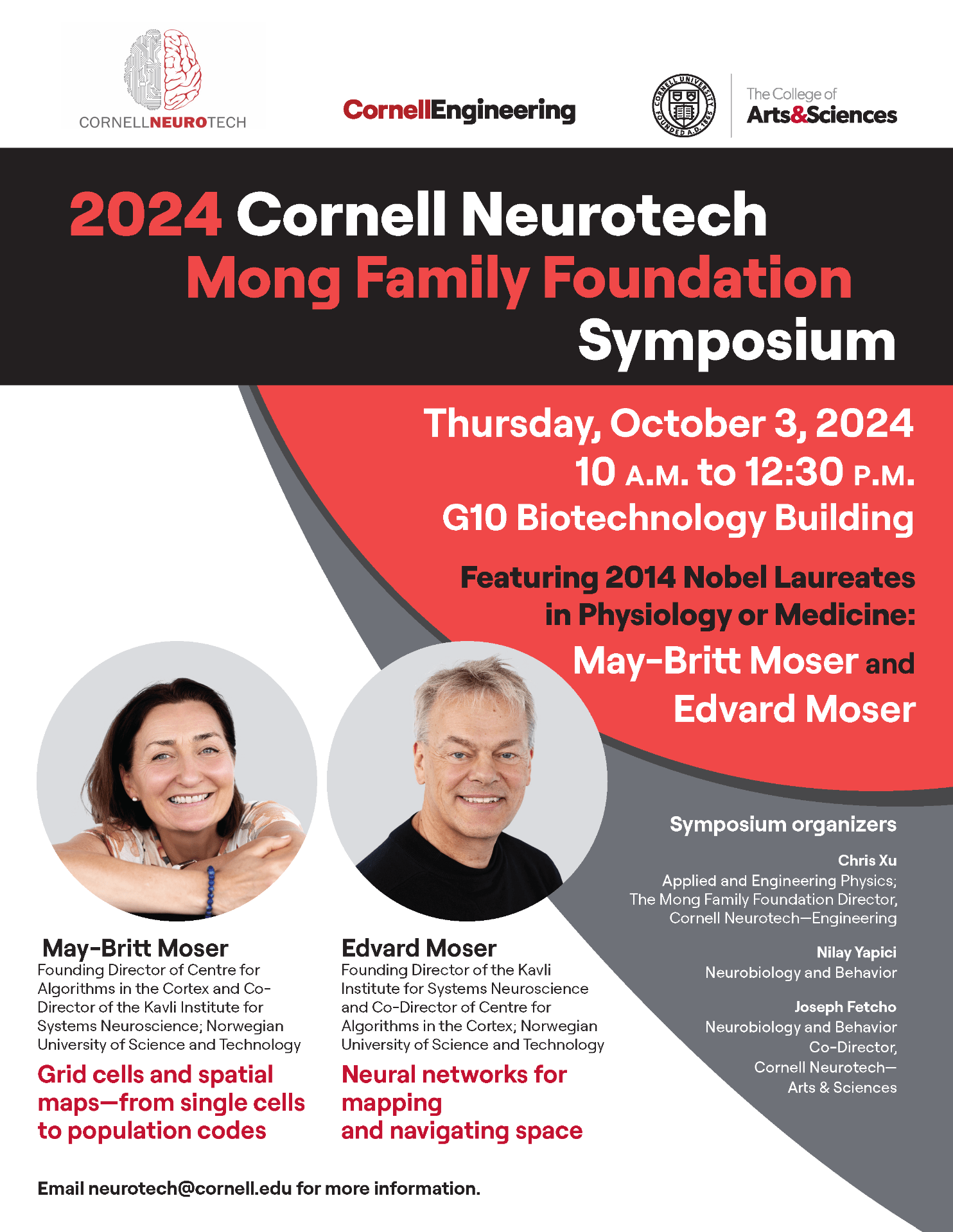
2024 Cornell Neurotech Mong Family Foundation Symposium featuring Nobel Laureates May-Britt Moser and Edvard Moser
Imaging of neural structure and activity in Drosophila through the intact cuticle published in eLife
Multi-color 3-photon deep imaging is published in Science Advances
New imaging technique sheds light on adult zebrafish brain
NeuroNex Technology Conference
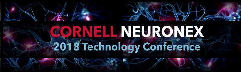
Wednesday, July 18 and Thursday, July 19, 2018
Cornell University, Ithaca, New York
The goal of the NSF NeuroNex initiative is to clear major technological hurdles in order to better study and understand the brain. Thank you to those who who made the 2018 NeuroNex Technology Conference possible. Please stay tuned for next year’s details. Co-organizers: Chris Xu, Joe Fetcho, Mert Sabuncu, Chris Schaffer, and Nilay Yapici. For details please visit: http://neuronex.cornell.edu
Gerald Rubin to speak at Cornell Neurotech seminar, Sept. 24, 2021 4PM
The annual Cornell Neurotech Mong Family Foundation Seminar will feature Gerald M. Rubin. The seminar will be held in G10 Biotechnology on Sept. 23, 2021 at 4pm. The seminar title will be:
Generating and using wiring diagrams of brain: Lessons from the Drosophila connectome
Gerald Rubin is a Senior Group Leader at the Howard Hughes Medical Institute (HHMI) Janelia Research Campus. He is the John D. MacArthur Professor of Genetics, Emeritus, at University of California Berkeley. His early work pioneered tools for germ line transformation using P-element transposons in the fruit fly Drosophila. This and other genetic tools allowed him to reveal signaling pathways downstream of tyrosine kinases. He went on to lead the project to sequence the fly genome. In recent years he has focused on revealing principles of how the brain gathers, stores and processes information. Rubin and his team develop advanced tools and methods to find, map, and analyze the neuronal circuits underlying learning, memory, sleep regulation, visual perception, and female-female aggression in Drosophila.
Early career scientists named 2017 Mong Fellows in Cornell Neurotech
Ten new Mong Family Foundation Fellows in Neurotech will work under the mentorship of faculty across Cornell to advance technologies that promise to provide insight into how brains work, as well as strategies to fix them when they don’t. Read the full Cornell Chronicle article here.
NeuroNex—a Radical Collaboration
An engineer and a neuroscientist gathered a group of Cornell scientists and engineers to tackle a frontier of science—the brain. Now, they form a hub.
by Jackie Swift, CornellResearch
Read the article and learn more here.
$9M grant will create neurotech research hub at Cornell
Aug. 1, 2017
By Syl Kacapyr for the Cornell Chronicle
As neuroscientists examine challenging questions about the complexities of the central nervous system, new tools to be developed at Cornell will provide them with an unprecedented glimpse into the inner workings of the brain thanks to a five-year, $9 million grant…Read the full article here.
3-photon imaging of mouse brain activity is published in Nature Methods
Dimitre G Ouzounov, Tianyu Wang, et al. record spontaneous activity from up to 150 neurons in the hippocampal stratum pyramidale at ~1-mm depth within an intact mouse brain.
Using three-photon microscopy at 1,300-nm excitation, the authors of a recently published Nature Methods paper, demonstrated that functional imaging of GCaMP6s-labeled neurons can be achieved beyond the depth of two-photon microscopy. The method creates opportunities for noninvasive recording of neuronal activity with high spatial and temporal resolution deep within scattering brain tissues.